
CT-BIT: CT Bone Investigational Toolkit
Get the best tools for understanding bone status, structure and change
Standard volumetric QCT can provide a detailed 3D representation of bone structure at the spine and hip. The Mindways CT Bone Investigational Toolkit (CT-BIT) extends our QCT Pro™ software to produce accurate BMD analysis beyond these regions. In addition, CT-BIT calculates mineral density distribution and geometry information to allow the characterization of bone status or change in response to therapy, aging or disease processes. Through its script-driven flexibility, the QCT Pro™ CT-BIT extension provides access to a vast array of measures and capabilities for the investigation of bone.
Volumetric pQCT
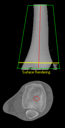
Until recently, pQCT has usually been performed on a dedicated machine, but there are significant advantages to performing pQCT bone density assessment on a clinical Whole Body Scanner using QCT ProTM CT-BIT:
- The cost of adding a QCT system to a standard CT scanner is considerably less than the cost of a dedicated pQCT machine;
- Patient motion artifacts are reduced with scan times of a few seconds instead of the 90 seconds per slice using a dedicated pQCT machine;
- Accurate matching of sections of bone during growth are feasible with a volume compared with scanning a few slices using a dedicated machine.
Cortical Thickness Measures
Focal thinning of cortical bone in the proximal femur may predispose a hip to fracture. Detecting such defects in clinical CT is challenging; cortical bone may be significantly thinner than the imaging system resolution. Accurate cortical thickness estimation requires good estimation of the cortical density which is provided by QCT.
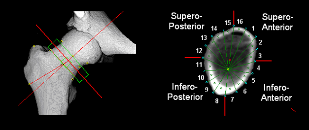
The CT-BIT extension provides a flexible script-driven framework for cortical thickness measurement using QCT data from QCT Pro™.
Comparison of measurements may be made using an automated frame of reference based upon anatomical landmarks for accurate registration.
In addition, cortical thickness and distribution measurement may be made at anatomical sites other than the hip.
HSA and Cross Sectional Analysis
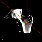
CT-BIT incorporates standard Hip Structural Analysis (HSA) measures using DXA-like area density projections methods to analyze three cross-sectional profiles at the Femoral Neck (FN); Intertrochanteric (IT) and Femoral Shaft (FS) regions. For each region, the distribution of the bone mass across the bone is extracted then parameters related to geometry and mechanical strength are calculated.
Volume density in cortical, trabecular and total bone compartments may also be extracted at the spine or peripheral sites such as the tibia or radius. In addition, CT-BIT includes template analysis scripts to help you customize and automate the complex analysis of your sets of QCT image data.
Data Explort
The QCT Pro™ Bone Investigational Toolkit Includes: